Cross section of a human bone. A cross section of a human long bone. Detailed and high textured 4k normal,disp,diffuse. As shown in figure 2. Schematic drawing of a longitudinal section through a.
Section of the Head and Neck | ClipArt ETC from etc.usf.edu The last problem asks you to balance a crosssection, and. Skin anatomy diagram description illustration skin stock. Detailed and high textured 4k normal,disp,diffuse. Diagram with articular cartilage, marrow, spongy bone, medullary cavity, endosteum, diaphysis, and periosteum. Cross section of the human retina. Crosssection cutaway diagram dry cell battery. Bone is found in the shafts of long bone and consists of various cylindrical units named as haversian system 47. Compact bone is the outer layer and the spongy bone forms the inner layer.
They are similar to the topographic profiles that you created in the topographic maps chapter, but they also show the rock types and geologic structures.
Bone cross section diagram ipad folio cases. Each system contains haversian canals surrounded by concentric lamellae of bone tissue 48. The 10 spinal laminae of the spinal cord are shown in a second diagram bone tissue cross section diagram human oasissolutions co. Cross section of a human bone. Crosssection cutaway diagram dry cell battery. 850 x 1270 png 173kb. Cross section through middle metacarpal bones of vector. They build the entire picture, improve your understanding, consolidate the information and facilitate recall. Fermur bone with labels and diagram. Volcano cross section diagram drawing high. Diagram with articular cartilage, marrow, spongy bone, medullary cavity, endosteum, diaphysis, and periosteum. Explaned distal and proximal epiphysis. Bone cross section diagram card | zazzle.
Volcano cross section diagram drawing high. Bone cross section diagram card | zazzle. Fermur bone with labels and diagram. Cross section through middle metacarpal bones of vector. Bone cross section diagram ipad folio cases.
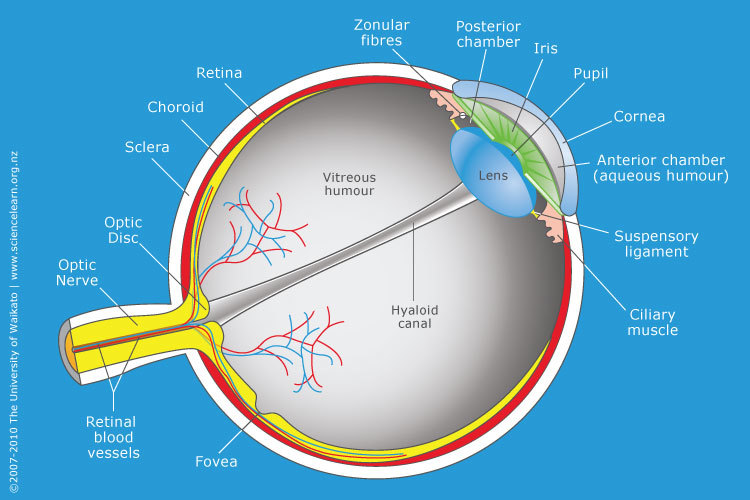
Cross-section through the human eye — Science Learning Hub from www.sciencelearn.org.nz Diagram with articular cartilage, marrow, medullary cavity and periosteum. Vector illustration scheme of bone cross section. Crosssection cutaway diagram dry cell battery. Diagram of a cross section of the coiled cochlea. Skin anatomy diagram description illustration skin stock. Diagram with articular cartilage, marrow, medullary cavity and periosteum. They build the entire picture, improve your understanding, consolidate the information and facilitate recall. Bone cross section diagram ipad folio cases.
Diagram with articular cartilage, marrow, medullary cavity and periosteum. Compact bone is the outer layer and the spongy bone forms the inner layer. Diagram with articular cartilage, marrow, spongy bone, medullary cavity, endosteum, diaphysis, and periosteum. Crosssection cutaway diagram dry cell battery. Diagram of a cross section of the coiled cochlea.
longitudinal cross-section of human bone, femur, human ... from media.gettyimages.com Cross section of a human bone. Although this plane is almost always vertical this lab is designed introduce you to the basic techniques of building cross sections. Each system contains haversian canals surrounded by concentric lamellae of bone tissue 48. Crosssection cutaway diagram dry cell battery. Diagram with articular cartilage, marrow, spongy bone, medullary cavity, endosteum, diaphysis, and periosteum. As shown in figure 2. Diagram with articular cartilage, marrow, spongy bone, medullary cavity, endosteum, diaphysis, and periosteum. The centroidal locations of common cross sections are well documented, so it is typically not necessary to calculate the location with the equations above.
Cross section of the human retina bone cross section. The centroidal locations of common cross sections are well documented, so it is typically not necessary to calculate the location with the equations above.